E-ISSN:2349-3275
P-ISSN:2349-5502

Case Report

Osteoma

Biomedical Review-Journal of Basic and Applied Medical Science

2018 Volume 5 Number 1 Jan-Dec
Publisherwww.biomedicalreview.in

A rare form of peripheral osteoma: Case report

Kumar L.K S.1*, Sudheena R.2, Menon.P V.3
DOI:

1* Surej Kumar L.K, Oral & Maxillofacial Surgery, Oral & Maxillofacial Surgery Kerala Institute of medical sciences (KIMS) Hospital, Trivandrum, Kerala, India.

2 R Sudheena, Oral & Maxillofacial Surgery, Oral & Maxillofacial Surgery Kerala Institute of medical sciences (KIMS) Hospital, Trivandrum, Kerala, India.

3 Varun Menon.P, Oral & Maxillofacial Surgery, Oral & Maxillofacial Surgery Kerala Institute of medical sciences (KIMS) Hospital, Trivandrum, Kerala, India.

Osteomas are slow growing benign osteogenic tumours with very low incidence rate. Though asymptomatic, sometimes the lesions may be large enough to cause cosmetic disfigurement and may need surgical excision. Peripheral osteoma of mandible is very uncommon and the one with nodules on the surface is extremely rare. We report an unusual form of peripheral osteoma with multiple nodules.

Keywords: Osteoma, Benign neoplasm, Peripheral type

Corresponding Author How to Cite this Article To Browse
Surej Kumar L.K, , Oral & Maxillofacial Surgery, Oral & Maxillofacial Surgery Kerala Institute of medical sciences (KIMS) Hospital, Trivandrum, Kerala, India.
Email:
Surej Kumar L.K, R Sudheena, Varun Menon.P, A rare form of peripheral osteoma: Case report. Biomed Rev J Basic Appl Med Sci. 2018;5(1):10-13.
Available From
http://www.biomedicalreview.in/a-rare-form-of-peripheral-osteoma-case-report

Manuscript Received Review Round 1 Review Round 2 Review Round 3 Accepted
2018-12-08 2018-12-10 2018-12-17 2018-12-24 2018-12-31
Conflict of Interest Funding Ethical Approval Plagiarism X-checker Note
Nil Nil Yes 17% IJB Archive

© 2018by Surej Kumar L.K, R Sudheena, Varun Menon.Pand Published by Siddharth Health Research and Social Welfare Society. This is an Open Access article licensed under a Creative Commons Attribution 4.0 International License https://creativecommons.org/licenses/by/4.0/ unported [CC BY 4.0].

Introduction

Osteoma a Latin term was first used in 1849, to denote a benign tumour composed of bone tissue.

Osteomas are benign osteogenic tumours of the craniofacial bones and may be central, peripheral or extraskeletal [1].

Of these, the solitary variety is a rare entity. Usually they are asymptomatic and slow growing, which may produce swelling and asymmetry. It is generally considered as a developmental anomaly and rarely as a true neoplasm [1].

Pathogenesis is unclear, but possibility of a reactive mechanism triggered by trauma or infection has been proposed [2], and also may be associated with certain syndromes [3].

Here we report an unusual case of peripheral osteoma in the angle of mandible with multiple projections on its surface.

Case report

A 42 year old male patient presented with a slow growing swelling on the right angle region of mandible with duration of more than two to three years.

It was asymptomatic and his major complaint was facial asymmetry and disfigurement. No history of trauma was elicited.

On examination, a bony hard swelling 3×3 cm was noted on the right angle region (Fig .1).

It was non tender. Skin over the swelling was smooth and non-pinchable, with small nodular lesions on the surface.

CT scan revealed a large pedunculated bony projection with multiple nodules on the surface (Fig 2a.2b).

A provisional diagnosis of peripheral osteoma was made and surgical excision was planned under general anaesthesia.

Through an extra oral approach, the lesion was exposed (Fig 3) and excised in toto (Fig 4) and smoothening of the bony margins was done (Fig 5).Haemostasis was achieved and primary closure was done.

ij_40_01.JPG
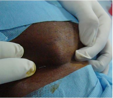
Fig 1: Clinical picture
ij_40_02.JPG
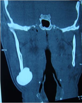
Fig 2a: Coronal CT scan showing the radioopaque mass
ij_40_03.JPG
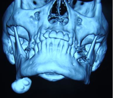
Fig 2b: 3D CT showing the attached mass to the mandible


ij_40_04.JPG
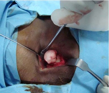
Fig3: Intra operative view showing the massij_40_05.JPG
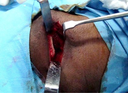
Fig4: Surgical site after excision of the lesion
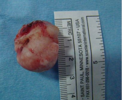
ij_40_06.JPGFig5: Excised mass

Discussion

The incidence of osteoma is comparatively very low, amounting to 0.01-0.04% of the population [3]. 22.85% lesions occur in the mandible and the incidence in maxilla is 14.2% [1].

2

The occurrence of osteoma is not age dependent, but commonly seen in young adults. Controversies exist for its gender predilection. Some authors believe that males are more affected than females in a ratio of 2:1 ([1], [2], [4]). But Bodner et al have stated that there is no predilection for age or gender [5].

The etiopathogenesis of osteoma is unclear. Traumatic, inflammatory, congenital and endocrine causes are considered as possible etiological factors [6].

The traumatic theory is the most accepted one. Supporting this theory, some authors have stated that the sites which are more susceptible to trauma, like lower border or buccal aspect of mandible have greater incidence of the lesion ([2], [5], [7]). Proximity to the muscle attachment suggests that muscle traction may have a role in its development ([1], [2]). We feel that minor trauma may have caused a subperiosteal haematoma, which combined with muscle pull may have initiated the formation of such lesions. It has also been reported in animal studies that Reilly- Finkel-Biskis osteoma virus (ecotropic type C retro virus) and the clones induce osteoma in mice [8].

Clinical appearance is usually round and smooth, solitary in nature ([5], [9]). Being asymptomatic, the discovery of peripheral osteoma is mostly an incidental finding. Diagnosis is based on clinical as well as radiographic appearance. Conventional radiographs like OPG or CT scan are used for diagnostic purpose. Three dimensional reconstruction provides the exact localisation and nature of the lesion [10].

We were able to identify multiple nodular projections on the solitary osteoma in 3 dimensional reconstruction, which is a very rare entity.

The differential diagnosis of peripheral osteoma includes exostoses, peripheral ossifying fibroma, periosteal osteoblastoma, osteoid osteoma and parosteal osteosarcoma ([1], [11]).

Treatment option is surgical excision and is indicated only for cosmetic reasons and rarely for functional impairment. Intra oral or extra oral approaches may be used. We went in for an extra oral approach since the lesion was extending towards the lingual aspect, as well as for the large size.


Conclusion

Peripheral osteoma of mandible is a rare lesion and multiple projections on the lesion, as seen in our case, is a very rare entity. We feel that recurrence rate is low after surgical excision, but it is advisable to have a long term radiographic follow up.

Reference

01. Sayan NB, Ucok C, Karasu HA, Guhan O. Peripheral osteoma of the oral and maxillofacial region: a study of 35 new cases. J Oral Maxillofac Surg 2002; 60: 1299-301. . [Crossref][PubMed][Google Scholar]

02. Kaplan I, Calderon S, Buchner A. Peripheral osteoma of the mandible: a study of 10 new cases and analysis of the literature. J Oral Maxillofac Surg 1994; 52:467-70. . [Crossref][PubMed][Google Scholar]

03. Singhal P, Singhal A, Ram R, Gupta R. Peripheral osteoma in a young patient : A marker for pre cancerous condition? J Indian Soc Pedodo Prev Dent 2012; 30:74-7. . . [Crossref][PubMed][Google Scholar] [Crossref][PubMed][Google Scholar]

04. Bosshardt L, Gordon RC, Westerberg M, Morgan A. Recurrent peripheral osteoma of mandible: report of case. J Oral Surg. 1971;29(6):446-50. [Crossref][PubMed][Google Scholar]

05. Bodner L, Gatot A, Sion-Vardy N, Fliss DM. Peripheral osteoma of the mandibular ascending ramus. J Oral Maxillofac Surg. 1998; 56(12): 1446-9. [Crossref][PubMed][Google Scholar]

06. Lucas RB. Pathology of tumours of the oral tissues. Edinburgh, Scottland: Churchill Livingstone; 1984. p. 191-4 [Crossref][PubMed][Google Scholar]

07. Schneider LC, Dolinsky HB, Grodjesk JE. Solitary peripheral osteoma of the jaws: A report of case and review of literature. J Oral Surg 1980;38: 452-5. . [Crossref][PubMed][Google Scholar]

08. Veera SD, Jayanthi K, Dayanand B. Solitary peripheral osteoma at a curious site with an ambiguous etiopathogenesis: A case report and review of literature. Int J Oral and Maxillofac Pathol 2012;3:50-5. . [Crossref][PubMed][Google Scholar]

09. Durighetto AF Jr, de Moraes Ramos FM, Rocha

MA, da Cruz Perez DE. Peripheral osteoma of the maxilla: report of a case. Dentomaxillofac Radiol 2007; 36:308-10. . [Crossref][PubMed][Google Scholar]

10. Bodner L, Bar- Ziv J, Kaffe I. CT of cystic jaw lesions. J Comp Assist Tomog 1994; 18:22-6. . [Crossref][PubMed][Google Scholar]

11. Longo F, Califano L, De Maria G et al. Solitary osteoma of the mandibular ramus: report of a case. J Oral Maxillofac Surg 2001; 59:698-700. . [Crossref][PubMed][Google Scholar]