E-ISSN:2349-3275
P-ISSN:2349-5502

Case Report

Cleidocranial Dysplasia

Biomedical Review-Journal of Basic and Applied Medical Science

2019 Volume 6 Number 1 Jan-Dec
Publisherwww.biomedicalreview.in

Cleidocranial Dysplasia: A Case Report

Kumar L.K S.1, Menon.P V.2*
DOI:

1 Surej Kumar L.K, MDS Consultant, Oral & Maxillofacial Surgery, Kerala Institute of medical sciences (KIMS) Hospital, Trivandrum, Kerala, India.

2* Varun Menon.P, Trainee, Oral & Maxillofacial Surgery, Kerala Institute of medical sciences (KIMS) Hospital, Trivandrum, Kerala, India.

Cleidocranial dysplasia (CCD) is a rare autosomal dominant disorder. This congenital defect is characterized by hypoplastic or absent clavicles, delayed skeletal development .Along with hypoplasia of orofacial and skeletal structures the condition is also reported to have multiple supernumerary teeth, retained deciduous dentition, non-eruption of permanent tooth. We report a case of CCD in a 26 year-old female who had classical diagnostic feature.

Keywords: Cleidocranial dysplasia, multiple missing tooth, absence of clavicles

Corresponding Author How to Cite this Article To Browse
Varun Menon.P, Trainee, Oral & Maxillofacial Surgery, Kerala Institute of medical sciences (KIMS) Hospital, Trivandrum, Kerala, India.
Email:
Surej Kumar L.K, Varun Menon.P, Cleidocranial Dysplasia: A Case Report. Biomed Rev J Basic Appl Med Sci. 2019;6(1):42-45.
Available From
http://www.biomedicalreview.in/cleidocranial-dysplasia-a-case-report

Manuscript Received Review Round 1 Review Round 2 Review Round 3 Accepted
2019-12-09 2019-12-10 2019-12-17 2019-12-24 2019-12-31
Conflict of Interest Funding Ethical Approval Plagiarism X-checker Note
Nil Nil Yes 18% IJB Archive

© 2019by Surej Kumar L.K, Varun Menon.Pand Published by Siddharth Health Research and Social Welfare Society. This is an Open Access article licensed under a Creative Commons Attribution 4.0 International License https://creativecommons.org/licenses/by/4.0/ unported [CC BY 4.0].

Introduction

Cleidocranial dysplasia (CCD) is a rare congenital autosomal dominant condition, usually presents with skeletal defects of multiple bones, the most characteristic feature of which are partial or complete absence of clavicles, wormian bones ,presence of open skull sutures and delayed closure of fontanels, and. These patients usually complain of multiple missing teeth or delayed eruption of teeth. Ours was a typical case of CCD in a 26 year old female patient who had classical diagnostic feature of this syndrome.

Case Report

A 26 year old female patient reported our department with complains of missing multiple teeth (Fig 1). Her family history showed no similar history. She was moderately built and nourished. Extraoral examination showed the patient had a flat face, hypertelorism and sunken nasal bridge. There was evidence of clubbing of the digits (Fig 2), shoulders appeared narrow and showed marked drooping. On palpation, there was bilateral absence of clavicles and patient was able to approximate both shoulders in the midline on the chest. Intraoral examination revealed the presence of both deciduous and permanent tooth with several missing tooth in a narrow constricted maxillary and mandibular arch.

Orthopantomogram (Fig 3) showed mandible with coarse trabeculation, narrow ascending ramus and slender pointed coronoid processes and multiple impacted tooth .The posteroanterior view of chest radiograph (Fig 4) revealed complete bilateral absence of clavicle. Based on the typical clinical examination and radiographic feature, a diagnosis of cleidocranial dysplasia was made.

ij_46_01.JPG
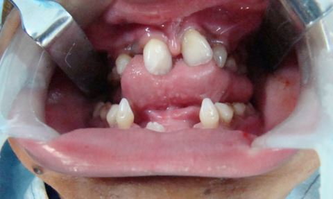
Fig 1: Showing the Intra oral Picture

ij_46_02.JPG
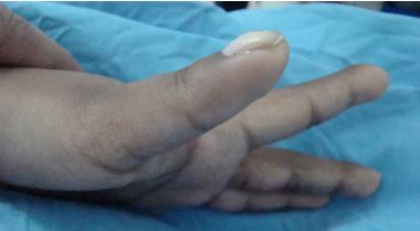
Fig 2: Clubbing of the digits
ij_46_03.JPG
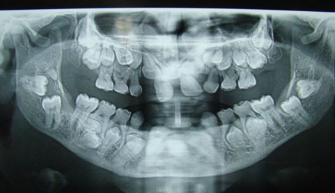
Fig 3: OPG showing multiple impacted tooth and retained deciduous tooth
ij_46_04.JPG
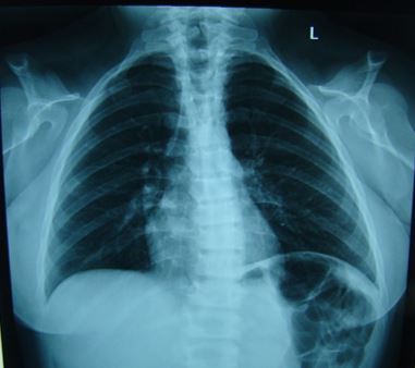
Fig 4: Chest X-ray (PA view) showing bilateral absence of clavicle

Discussion

Cleiodocranial dysplasia (CCD), also known as Marie and Sainton’s disease; Scheuthauer - Marie – Sainton syndrome; Mutational Dysostosis and cleidocranialdysostosis [1].

CCD is generally accepted as an autosomal disorder, affecting both sexes with equal frequency. In 1765 CCD was first described by Martin [2] and in 1897 Marie and Sainton termed


it as ‘cleidocranial dysostosis’ [3]. The aetiology of cleidocranial dysplasia is unknown but Genes has been mapped for this kind of disorder ([4, 5]).

CCD is usually seen involved with the bones that get ossified early in foetal life; the clavicle is the first bone to show ossification. The disease characteristically causes a retardation or partial aplasia of bones that ossify intramembraneously ([4, 6]). It is rather an uncommon disorder with a prevalence of 0.5 per 100,000 live births [7]. The condition is characterised by abnormalities of the clavicles, skull and jaws as well as by occasional stunting of long bones. The patient is usually of short stature, but the neck appears relatively long and the most characteristic and differentiating skeletal feature is that of partial or completely ab­sence of one or both clavicles and drooping of shoulders can be seen as a result of this. Similar features were seen in our case.

The intraorally the condition is characterised by the presence of multiple supernumerary teeth and the delayed eruption of the succedaneous dentition [8]. The middle third of face is usually hypoplastic both in the antero-posterior as well as in vertical dimensions. Hard palate is high and narrow. The causes of unerupted teeth in CCD is said to be [9] (i) a disturbance of bone resorption and delay in formation of tooth and its maturation (ii) delay and defectiveness in formation of cellular cementum or (iii) interposed fibrous tissue acting as a barrier to eruption resulting in a non union between the dental follicle and the mucosa.

The diagnosis is confirmed by radiographs [10]. Chest radiograph in these patients shows hypoplastic or aplastic clavicle. Radiographically, the mandible and maxilla contained many unerupted teeth that are greatly deviated from the normal position [10] similar features were reported in our case.Crane- Heise syndrome, mandibuloacral dysplasia, pyc­nodysostosis, yunis varon sydrome, CDAGS syn­drome and hypophosphtasia etc. are usually considered  in the differential diagnosis of CCD [11].

There has been no specific treatment for the patient with CCD; the correction of the dentofacial deformities both aesthetically and functionally has been mentioned in literature. Most of the times the age of the patient determines the treatment plan; in the early age group, the supernumerary teeth are

surgically exposed and orthodontic traction is applied with the help of brackets until the teeth are brought into normal occlusion [12].Orthognathic surgery has been suggested by Trimble et al, [13] for the correction of middle - third of the face. Removable prosthesis has been advocated by Winter after the extraction of all the primary and permanent teeth [13] but these patients most of the time ends up with alveolar hypoplasia which compromises the retention of prosthesis. Suggested removal of only the erupted teeth and use of a removable prosthesis inorder to minimize the alveolar bone loss was proposed by Pusey & Durie [14].It is not always easy to determine the most appropriate aesthetic and functional treatment for dentofacial deformities in patient with CCD ,so the ideal treatment for these patients still remains a myth.

Conclusion

A multidisciplinary approach to management of dental abnormalities on a long-term basis is necessary in the treatment of CCD [15]. Early diagnosis of CCD ensures an aesthetic facial appearance, functional occlusion and better quality of life for the patient.

Reference

1. Shafer WG, Hine MK, Levy BM. A text book of Oral Pathology,4th ed. Philadelphia, WB Saunders Co. 1993;678-80.

2. Martin M. Sur undeplacement natural de la clavicule. J Med ChirPharmacol. 1765; 23 :456.

3. Marie P, Sainton P. Observation d’hydrocephalic hereditary (pereetfils), par vice de development du crane et du cerveau. Bull Soc Med Hop Paris.1897; 14:706.

4. Mundlos S, Mulliken JB, Abramson Dl, Warman ML, Knoll JHM & Olsen BR. Genetic mapping of cleidocranial dysplasia and evidence of a microdeletion in one family. Hum Mol Genet.1995;4:71-75.

5. Mundlos S. Cleidocranial dysplasia: clinical and molecular genetics. J Med Genet. 1999; 36(3):177-82.

6. Currall V, Clancy R, Diamond D. Cleidocranial dysplasia. Curr Orthop. 2007; 21(2): 159-62.


7. Yochum TR and Rowe LJ. Essentials of skeletal Radiology, Williams & Wilkins, Baltimore, Md, USA, 2nd Edition, 1996.

8. Yamamoto H, Sakae T, Davies JE. Cleidocranial Dysplasia: A light microscope electron microscope and crystallographic study. Oral Surg Oral Med Oral Path. 1989; 68: 195 - 200.

9. Amita Sharma, Sansar Sharma. Cleidocranial Dysplasia - Report of A Case.J Oral Health Comm Dent 2009;3(3)62-65

10. Koch PE, Hammer WB. Cleidocranialdysostosis: review of literature and report of case.J Oral Surg. 1978; 36: 39-42.

11. Dixit R, Dixit K, Paramez AR. Cleidocranial dysplasia. Lung India 2010; 27 (3):176-7.

12. FerdaTasar, EnginBelut, Celal Turner, Mustafa Saysel, Mehinet Muhtarogullan. Clediocranial dysplasia: case report. Austr Dent J. 1995; 40(6): 352-56.

13. Trimble LD, West RA, McNeill RW. Cleidocranialdysplasia. Comprehensive treatment of thedentofacial abnormalities. Jam Dent Assoc. 1982; 105: 661-6.

14. Pusey RF, Durie JF. A case of cleidocranialdysostosis showing failure of eruption of teeth. Br Dent J. 1943; 75(1): 11-13.

15. Ozkan G, Kanli A. Clinical and radiological manifestations of cleidocranial dysplasia: A Case report. Indian Journal of dentistry. 2013; 4(3):174 -77.